Molemate SIAscopy is a non -invasive, quick and pain free treatment. The MoleMate SIAscopy is placed over the lesion and a picture of it comes onto the screen. The process uses non-harmful light and digital imaging to evaluate the patient’s skin. The practitioner can then accurately detect melanoma at an early and treatable stage, dramatically reducing the need for biopsies and excisions of moles that are not suspicious.
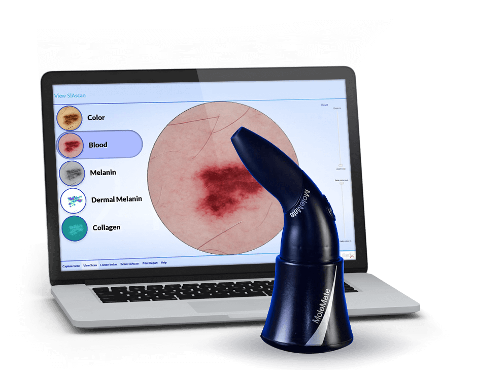
MoleMate SIAscopy is the only technology that can look 2mm beneath the skin’s surface to provide five distinct images to aid our diagnosis:
MoleMate SIAscopy reveals the position, spatial distribution, and concentration of, epidermal pigment, dermal pigment, haemoglobin, and collagen, representing important and critical markers to determine the state or condition of the mole or lesion.

What technology does MoleMate utilize?
MoleMate uses SIAscopy™, which is a clinically proven, non-invasive skin-imaging technology that can accurately help physicians detect melanoma at an early and treatable stage, dramatically reducing the need for biopsies and excisions of moles that are not suspicious. The process uses non-harmful light and digital imaging to evaluate the patient’s skin.
What does the word SIAscopy mean?
Spectrophotometric Intracutaneous Analysis, and utilizes a handheld LED-Light based spectrophotometer. SIAscopy projects visible and infra-red light to create an image of key components just beneath the patient’s skin.
SIAscopy reveals the position, spatial distribution, and concentration of, epidermal pigment, dermal pigment, haemoglobin, and collagen, representing important and critical markers to determine the state or condition of the mole or lesion.
Get in touch to book an appointment